
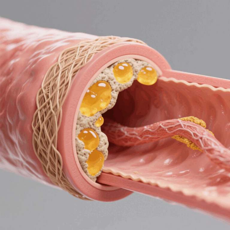
Labs planning next-year preclinical work are making concrete choices about model fidelity and throughput — and the split between orthotopic and subcutaneous approaches is coming into sharper focus. In practical terms, teams balancing timeline, cost, and translational relevance often cross-reference data from metabolic disease models early in protocol design to align pharmacokinetics and tumor microenvironment expectations. Around research hubs in the San Francisco Bay Area, investigators routinely compare orthotopic xenograft outcomes to subcutaneous implants to shape dosing strategies and PD biomarker selection.
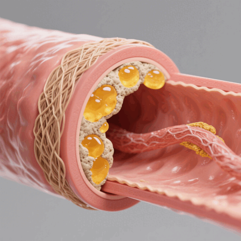
Comparative snapshot: what each model actually buys you
Subcutaneous models are fast and reproducible. Implant, measure calipers, run a growth curve — throughput and simple tumor-volume readouts are the advantage. Orthotopic models, by contrast, place tumors where they belong anatomically, so you capture relevant stroma interactions, invasion patterns, and sometimes metastatic routes. That realism improves translational signal for therapies that interact with microenvironmental cues — but it increases surgical skill needs, imaging complexity, and variability.
When to pick one over the other — clear trade-offs
Use subcutaneous when early screening demands scale: high cohort sizes, clear dose-response, and straightforward tumor burden metrics. Choose orthotopic when mechanism-of-action depends on local signals — immune infiltration, ECM remodeling, or organ-specific pharmacokinetics. For immuno-oncology or drug-conjugates that rely on tissue-specific uptake, orthotopic models reduce false negatives. Keep in mind xenograft source matters too: cell-line derived versus PDOs (patient-derived organoids) changes predictive power.
Emerging practices and humanized receptor models
Humanized receptor models — for example those incorporating GLP-1R and GIPR expression — are moving from boutique projects toward routine options for metabolic-oncology intersections. Integrating an in vivo metabolic disease model humanized gipr glp1r into an orthotopic workflow helps teams test dual-action agents on both tumor and systemic metabolic endpoints. Imaging, liquid biopsy PD biomarkers, and targeted pharmacokinetics sampling are the tools that make that integration meaningful. Teams in translational groups now treat receptor-humanized models as standard variables during go/no-go gate reviews — which reduces ambiguity later on.
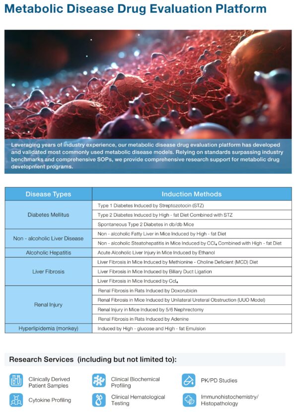
Operational production teardown: design, common mistakes, fixes
Operationally, the teardown starts with clear endpoints and sampling windows. Mistakes usually stem from fuzzy endpoints, underpowered cohorts, or mismatched PK sampling — and the fixes are straightforward: align assay sensitivity to expected concentrations, standardize surgical technique, and lock imaging schedules. Treat {main_keyword} and {variation_keyword} as operational parameters: define acceptable variance, set SOPs for implantation, and record environmental covariates. Small deviations in perioperative care skew growth rates — so standard operating steps pay off in reproducibility.
Vendor and platform selection: practical criteria
Compare vendors on three concrete axes: surgical proficiency for orthotopic implants, validated readouts (bioluminescence, MRI, histology scoring), and experience with humanized receptors like GLP-1R/GIPR. Ask for performance data from nearby sites — real lab service footprints around major hubs give you faster turnaround and peer benchmarking. — Also request raw endpoint distributions, not just mean curves; that reveals variability you’ll manage downstream.
Three golden metrics for selecting models and partners
1) Translational concordance: proportion of preclinical endpoints that matched early clinical PK/PD within an acceptable margin across prior projects. Track this as a percent over historical runs.
2) Endpoint robustness: coefficient of variation for primary tumor measures and key PD biomarkers across biological replicates. Aim for CVs under set thresholds tied to power calculations.
3) Operational reproducibility: percent of studies delivered on SOP timelines without protocol deviations. That metric predicts internal workload and downstream costs.
Teams that measure these three things sooner spend less time chasing artifacts and more time optimizing lead molecules. The practical result: clearer go/no-go decisions and fewer surprises when moving to IND-enabling studies. Final thought — for hands-on translational groups looking to blend metabolic receptor biology with tumor models, the pragmatic advantage is obvious: a partner who understands orthotopic nuance and receptor-humanization reduces repeat runs. Jennio Biotech. —